



OLYMPUS cellSens는 독특한 GUI (Graphic al User Interface)를 통해 샘플 이미지를 순차적으로 쉽게 사용할 수 있는 올림푸스 만의 이미지 프로그램입니다. 유저에 맞게 기본부터 최고의 사양 까지 선택하실 수 있으며 강력한 분석 툴을 제공합니다.

| 사용자를 위한 편의성 | ||
| 올림푸스의 새로운 cellSens software는 이미지를 촬영하고, 분석하는 흐름을 사용자가 원하는 대로 재배치 할 수 있습니다. | 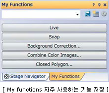 |
|
| My Functions: 사용빈도가 높은 창을 통해 별도 관리, 저장이 가능합니다. Process manager: 기능을 통해 전동현미경과 연동해 multi-color, timelapse, XY multi-position, Z-stack images를 손쉽게 촬영할 수 있으며 여러 기능들을 조합, 하나의 버튼으로 실행할 수 있습니다. Arrange windows: 사용자가 원하는 기능을 창안에서 배열이 가능합니다. |
||
 |
||
| cellSens 기능 | ||
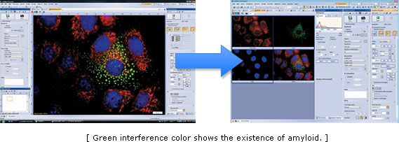
| Multi-dimensional Image XY(2D)이미지 부터 XYZ, Time, Multi color, Multi area(6D)이미지 까지도 다양하게 프로그램화 하실 수 있습니다. |
||
 |
||
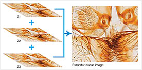
| EFI (Extended Focus Image) | ||
| 초점 심도를 달리하며 이미지를 찍고 합쳐 한 장의 또렷 한 사진을 만들거나 넓은 면적의 시료를 부분 별로 찍어 한장의 파노라마 사진으로 만들 수 있습니다. |
 |
|
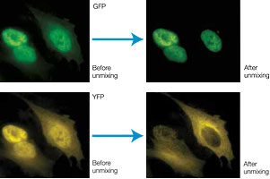
| Unmixing | ||
| 기능을 통해 emission spectra가 겹쳐 발생하는 Cross-talk 현상을 제거해 줄 수 있으며, 백그라운드의 자가형광 또한 분리할 수 있습니다. |
 |
|
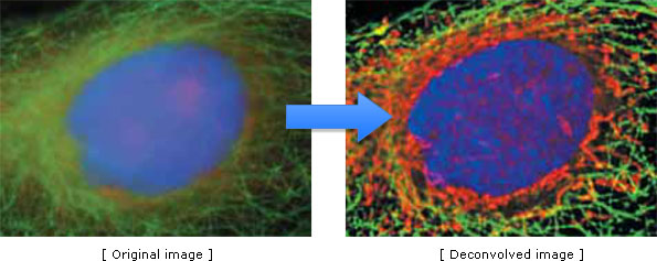
| Deconvolution Live 2D dconvolution algorithm은 이미지 미리보기를 이용해 더 좋은 focusing의 이미지를 촬영할 수 있게 해줍니다. 또한 deblurring 기술을 이용, 초점심도 밖의 data를 삭제하여 또렷한 이미지를 구현하며 CI deconvolution module을 채택 resolution, contrast, dynamic range를 증가 시켜 줍니다. |
||
|
|
||
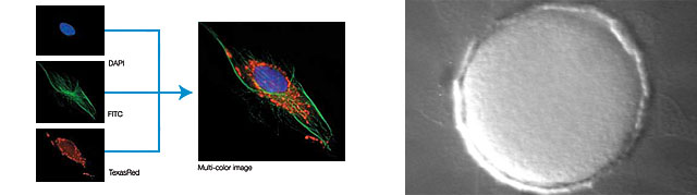
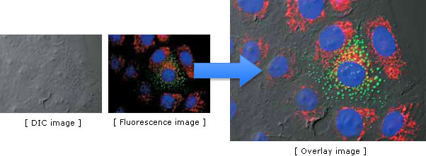
| Image Composition 이 편리한 기능은 다른 관찰법이나 다른 파장의 촬영된 이미지들을 한장의 이미지로 통합해 줍니다. 예를 들어 cell의 morphology와 localization을 관찰하기 위해 DIC 이미지와 형광 이미지들을 겹쳐 보길 원할 때 사용할 수 있습니다. |
||
|
|
||
| Image Enhancement Edge enhancement, smoothing, sharpening, shading correction, contrast control, DCE (Differential Contrast Enhanceme nt) 등의 다양한 filtering 기능을 지원합니다. cellSens software의 가장 낮은 Entry 등급에서도 simple-measurement 기능 을 이용할 수 있으며, 등급에 따라 point position, distance, angle, polygon area, boundary area, count, intensity 등 다양한 분석 기능이 가능합니다. |
||
 |
||
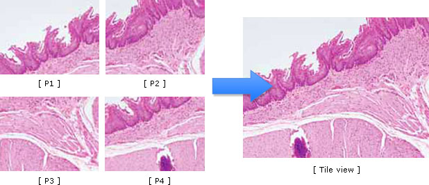
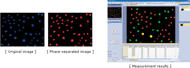
| Image Stitching 고배율의 이미지를 한 장에 합쳐 전체적인 이미지를 볼 수 있는 기능입니다. 뇌조직 이미지 및 렌즈에 담을 수 없는 부분의 영역을 한 장에 표현 가능합니다. Auto counting 세포개수 가운트, 면적 측정, 형광세기 측정을 쉽고 간편하게 하실 수 있습니다. 이러한 기능을 통해 다양한 분석부터 측정까지 완벽하고 정확한 값을 얻으실 수 있습니다. |
||
 |
||
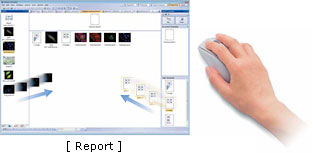
| Report cellSens software에서 생성된 자료는 Report 기능을 이용해 빠르고 쉽게 MS Excel의 데이터로 만들 수 있습 니다. |
 |
|
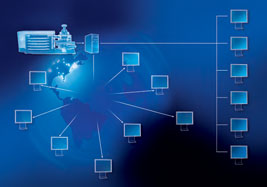
| Database (Global Discussion) SQL Server를 이용하여 이미지와 데이터를 다른 사용자 와 공유하고 저장하실 수 있어 장소와 시간의 상관 없이 어느 누구나 편리하게 자료를 사용하실 수 있습니다. |
 |
|
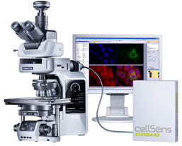
| Various Devices 올림푸스 현미경, 카메라 외 Hamamatsu, Q-Imaging, R oper, Jenoptik 카메라와 Sutter, Uniblitz, Prior, Ludl, Märzhäuser 등의 다양한 제품 컨트롤이 가능합니다. |
 |
|

Device control
| Dimension | Standard | Entry | |||
|---|---|---|---|---|---|
| Olympus | Camera | DP20*, DP21, DP25, DP26, DP70*, DP71, DP72 |
√ | √ | √ |
| Microscope | BX43, BX53, BX63, BX61, BX61WI, IX81, SZX16A | √ | √ | ||
| IX81-ZDC, IX81-ZDC2 | Multichannel 5D | ||||
| Peripherals | BX-DSU, IX2-DSU, BX-FCB, IX-FCB |
Multichannel 5D | |||
| Hamamatsu | Camera | Orca R2 (C10600-10B), Orca 03 (C8484-03G), Orca 05 (C8484-05G), ORCA ER (C4742-95-12ER), Orca Flash 2.8 |
√ | √ | |
| Q-Imaging | Camera | MicroPublisher 3.3 RTV, MicroPublisher 5 RTV |
√ | ||
| Exi Blue, RETIGA (Exi, SRV, 2000R, 2000RV, 4000R, 4000RV) plus RGB slider |
√ | ||||
| Photometrics | Camera | CoolSNAP HQ2 | √ | ||
| Jenoptik | Camera | ProgRes C3, ProgRes C5 | √ | √ | |
| Vincent Associates |
Shutter | Uniblitz shutter (VCM-D1, VMM-D1, VMM-D3) |
√ | √ | |
| CoolLED | Light Source | precisExcite (pE-1, pE-2) | √ | ||
| EXFO | Light Source | Lambda DG4 | √ | ||
| Sutter | Shutter | Uniblitz shutter (VCM-D1, VMM-D1, VMM-D3) |
√ | ||
| Prior | Motorized XY stage |
ProScan (I, II) | Multiposition | ||
| Shutter, FW, Z-drive |
ProScan (I, II) | √ | |||
| Ludl | Motorized XY stage |
MAC6000 | Multiposition | ||
| Shutter, FW, Z-drive |
ProScan (I, II) | √ | |||
| Objective Imaging |
Motorized XY stage controller |
Oasis 4i | Multiposition | ||
| Z-drive controller | Oasis 4i | √ | |||
| Marzhauser | Motorized XY stage |
Tango | Multiposition | ||
| * DP20/70 does not support Windows 7 64bit | |||||
Compatible image formats
| Read and write | VSI (Virtual slide image), JPEG, JPEG2000, TIFF, BMP, AVI, PNG |
|---|---|
| Read only | GIFF, PSD (Adobe PhotoShop), TIFF (DP-BSW, FSX100, MetaMorph), OIF/OIB (Fluoview format),Cell, STK (MetaMorph), MRC (Medical Research Council) |
System requirements
| OS | Microsoft Windows 7 (32-bit/64-bit) Ultimate Microsoft Windows 7 (32-bit/64-bit) Professional Microsoft Windows Vista (32-bit) Ultimate with SP2 Microsoft Windows Vista (32-bit) Business with SP2 Microsoft Windows XP Professional (32-bit) with SP3 |
|---|---|
| OS Language | English, Simiplified Chinese, Japanese and all others with English like alphabet |
| CPU | Intel Pentium® 4, Intel Xeon®, or Intel Core™ Duo (or equivalent) |
| RAM | 3 GB or more recommended (minimum 2 GB required) |
| Display | 1280x1024 or larger recommended (1024 x768 or larger), 32-bit video card |
| Port | USB port for protection key Serial (RS232) for hardware control (BX61, IX81, SZX2-MDCU etc...) |
| HDD | 1 GB or more of free space (at the time of installation) |
| Drive | DVD-ROM drive |
| Web Browser | MS IE 6.0 or later |
cellSens functions
| Dimension | Standard | Entry | ||
|---|---|---|---|---|
| Layout | User Experience Customization | √ | √ | √ |
| View | Overlay multiple images | √ | √ | |
| Document groups for side-by-side image comparison |
√ | √ | √ | |
| Movie playback | √ | √ | √ | |
| Tile View (multiple images in a single data set shown side by side) |
√ | √ | √ | |
| Slice View for orthogonal plane viewing of 3D or timelapse data sets |
√ | |||
| Voxel Viewer for isosurface and volumetric rendering of 3D and 4D data sets |
CI Deconvolution | |||
| Image Acquisition |
Snap / Movie Acquisition | √ | √ | √ |
| Time-Lapse at specified interval | √ | √ | ||
| Automated Multi-wavelength | Multichannel 5D | |||
| Z-Stack | √ | |||
| Multi-Dimensional (xyzt and wavelength) |
Multichannel 5D | |||
| Multiple Image Alignment (panorama) based on live image (manual) |
√ | |||
| Multiposition visitation and Stage Navigator |
Multiposition | |||
| Automated Multiple image Alignment (requires motorized stage) | Multiposition | |||
| Instantly create EFI image (manual or motorized Z) |
√ | |||
| Live deblurring | √ | |||
| Image Processing |
Geometry/combine/filter processing | √ | √ | |
| Fluorescence unmixing | Multichannel 5D | |||
| Brightfield unmixing | Count & Measure | |||
| Deblurring (No/Nearest Neighbor, Wiener Filter) |
√ | |||
| 3D Deconvolution (Constrained Iterative Deconvolution) |
Multichannel 5D | |||
| Image Analysis | Region and line measurements, | √ | ||
| Phase analysis | √ | |||
| Object analysis and classification | Count & Measure | |||
| Line profile | √ | √ | √ | |
| Intensity plot over time/z | √ | |||
| Colocalization | Multichannel 5D | |||
| Documentation and Collaboration |
Automatically compose Word reports | √ | ||
| Database image and data management solution for microscopy |
Database | |||
| * Three Points angle, four Points angle, Arbitrary Line, Closed polygon, Polyline and Perpendicular line only. | ||||